What to expect when you have a mole removed
If you have an abnormal mole, you may need to have it removed. Removing a suspicious mole and examining it for cancer cells is an important step in catching melanoma early, when it’s easiest to treat. During a skin cancer screening exam your dermatologist may find an abnormal mole. An abnormal mole could be a melanoma symptom, or it could be benign, meaning it’s not cancerous. To determine what type of cells make up the mole, the dermatologist will remove the mole for a biopsy.
Here’s what to expect if you are visiting the dermatologist.
Skin self-check
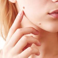
Before your appointment, you should check out your own moles by doing a skin self-exam. Look for the ABCs of melanoma, and note any of the following symptoms so you can point them out to your dermatologist. These skin cancer symptoms include:
- Asymmetry: The two sides of the mole look different from each other.
- Border: The mole’s border is crooked, jagged or irregular.
- Color: The mole is multi-colored.
- Diameter: The width is more than 6 millimeters, which is about the size of pencil eraser.
- Evolution: The mole has changed in size, shape or feeling.
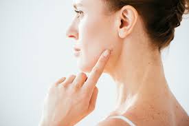
Dermatologist exam
During the appointment, your dermatologist will look for any abnormal moles. If an abnormal mole is noted on exam, your dermatologist may recommend monitoring it closely for any changes or removing it for a biopsy.
If a mole looks concerning, a biopsy is done so that the mole can be examined further under a microscope. This gives us a more definite diagnosis based on a close-up view of how the cells in the mole look and are arranged.
Removing a mole
First, the dermatologist will give you a numbing injection near the mole. This may pinch a little, but should keep you from feeling any pain during the removal. There are a few different techniques your dermatologist may use to remove the mole. These techniques include:
- Shave biopsy – a razor blade is used to shave off the mole and the skin around it
- Punch biopsy – A punch tool is placed over the mole and used to “punch” out the mole
- Scalpel removal – A scalpel is used to remove the mole and skin surrounding it and stitches are used to help the skin heal
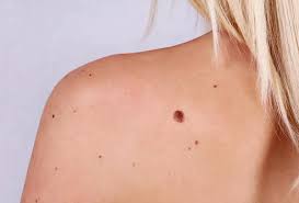
Normal vs. Atypical Moles
“Normal” moles are typically smaller than a pencil eraser, round and symmetrical with an even color and smooth borders. They are often tan or pink, and though you may not like them cosmetically, there is no medical reason to have them removed.
Atypical moles — also called dysplastic nevi — are unusual looking moles that are non-cancerous. When viewed at the cellular level, these moles look different than healthy, normal moles. They may be asymmetric, very dark or multi-colored and may have indistinct borders. Often found on the back, chest, buttocks or scalp, these moles look different than the other moles on your body.
Atypical moles may look like melanoma — the deadliest form of skin cancer — but don’t worry, they’re not cancerous. Atypical moles won’t necessarily develop into skin cancer, but if you have 10 or more dysplastic nevi moles, you do have a higher risk of developing melanoma (roughly 12 times the risk of the general population). Melanoma can spread very quickly but is curable if caught early.
How moles are removed
A mole can usually be removed by a dermatologist in a single office visit. Occasionally, a second appointment is necessary.
The two primary procedures used to remove moles are:
- Shave excision. For this procedure, your dermatologist uses a thin, razor-like tool to carefully slice away the mole. A device with a tiny electrode at the end may be used to perform electrosurgical feathering.
The feathering helps minimize the appearance of the excision by blending the edges of the wound with the surrounding skin. Stitches are not needed after a shave excision. The mole is usually examined under a microscope afterwards to check for signs of skin cancer.
- Surgical excision. This procedure is deeper than a shave excision and more like traditional surgery. Your dermatologist cuts out the entire mole and below to the subcutaneous fat layer, and stitches the incision closed. The mole will then be examined for cancer cells.
You should never try mole removal yourself. The risks of infection and bad scarring are too great. And if the mole was cancerous, you may leave cancer cells behind.
Self-checking moles
While they generally do not pose a health threat, moles should be self-checked regularly. Schedule a skin exam if you find a new mole or a change in an existing one. A mole that starts to change or grow in size could be a sign of skin cancer.
Whether you choose to have a mole removed for health or cosmetic reasons, it’s important to know what you’re dealing with first. It may seem convenient to use over-the-counter products or home remedies to remove small moles. But it is wise to check with your doctor to make sure the mole is not potentially cancerous.
Skin cancer screening:
If a mole starts to grow, itch, or bleed, make an appointment to see a dermatologist.
Moles are common. Almost every adult has a few moles. Adults who have light skin often have more moles. They may have 10 to 40 moles on their skin. This is normal.
You should not be overly worried about your moles. But you should know:
- A type of skin cancer, melanoma, can grow in or near a mole.
- Caught early and treated, melanoma can be cured.
- The first sign of melanoma is often a change to a mole — or a new mole on your skin.
- Checking your skin can help you find melanoma early. A dermatologist can show you how to examine your skin and tell you how often you should check your skin.